Sunday, November 10, 2013

The Reality of Virtuality

            One of the reasons it’s been a few weeks since a post is a recent Philosophy of Mind essay that has been consuming most of my free time. My chosen topic; If you were uploaded to a computer, would you retain your youness?  After doing some deep soul-searching for this essay, I was delighted to hear CBC’s program Spark include a segment of Virtual Reality last week, and the physical possibility of it. The segment (which I 
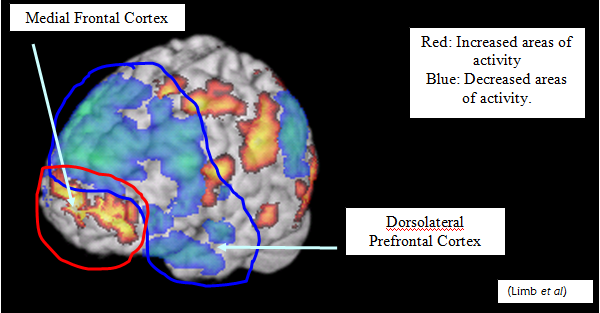
will link to at the bottom of the page) is an interview with Jody Culham, a professor at the department of psychology at Western University. Her research focus’ on how the brain processes real objects versus images of those objects. Culham’s research has shown that we process real objects very deeply. It is believed the reason for this does not have to do with the 3D vs 2D problem, but with possibility of interacting with the object.
            This is shown in a genius study by Antonio Ranguiel at CalTech. He brought in hungry students and gave them each money that they could bid with. He then presented them with an auction of different food. The variable was how the food was presented, some students were shown simply the name of the food, some a picture, some the food behind Plexiglas and some the actual food. Students were willing to spend 50-60% more on real food than any of the presentations, even the food behind the Plexiglas  which was at the same level as the pictures of the food. This supports the idea that it’s not just that we can see the 3D-ness of the object. It’s that we could reach out and touch the food if we wanted to.
            So is there any hope in achieving the perfect visual representation of something? Culham doesn't think so. “Some of the success of the more appealing [virtual] products is actually that they haven’t only focused on making very slick displays, but that they have focused on enabling the user to interact with them in a very intuitive way.” Think touch screens, wii games. We are interacting with these things as we would if they were real objects. It’s not the images we care about, it’s the level of interactions we can have with them.
            How about virtual reality. Even with the lack of object-realness, if the interactions are the same, could it work? Could we live in a matrix-like world? This time, Culham is optimistic. “Presumably as far as the brain is concerned as long as the inputs and the outputs are the same then we shouldn't be able to tell the difference between reality and virtual reality.”
            Imagine this future world: Babies would be genetically engineered and grown in labs, and taught by teacher robots. At the peak age, the brain would be mapped and a functionally equivalent program would be made. The program is turned on, and your new conscious life as a computer program begins. Your body would be disposed of, and you continue to live indefinitely in a computer-simulated reality. We hijack Mother Nature’s work, and improve it, make it last longer.             
             Would you want to live in this world? Would the interactions with virtual objects be enough? Would the interactions with virtual people be enough? Or would you still be missing that something?

Radio Segment on Spark: http://www.cbc.ca/player/Radio/Spark/ID/2414455201/
Jody Culham's Lab: http://culhamlab.ssc.uwo.ca/culhamlab/

Antonio Ranguiel's Lab: http://www.rnl.caltech.edu/

Saturday, October 12, 2013

Stopping the Shaking: Treating Parkinson's with Deep Brain Stimulation

          In June of this year, a video came out on Youtube that has now achieved over 1 million views. Unlike may viral Youtube videos, it is not images of cats chasing lasers, or people running into glass doors. Instead, it is of a man, sitting down in a chair talking to the camera. This man’s name is Andrew Johnson (AJ), and 4 years earlier at 35, just starting a family with 5 and 3 year old children, he was diagnosed with Early Onset Parkinson’s Disease. You would not know this by looking at the video though. Parkinson’s Disease is a progressive disorder affecting motor movement, most famously tremors and stiffness/slowing. The AJ on camera’s movements are fluid, his speech is normal and understandable. He goes through some simple motor tests with his hands, showing complete movement. AJ then goes on to explain in 2012, he underwent surgery for Deep Brain Stimulation (DBS). He picks up a remote control, puts it to his chest, and pushes a button. Almost immediately, his hand starts to tremor. He holds his hands up and they blur in front of the camera. His neck tightens and his head tilts. His speech is slow and broken. With trouble, he grabs the remote again and turns his device in his chest back on. Again, almost immediately, he is returned to his original state.
            At first watch, it is hard to believe. It seems that science has founds a complete cure for Parkinson’s. This is not the case though. DBS does not stop Parkinson’s symptoms as seen when AJ stops his stimulator, and it is often used alongside other treatment methods. It does not stop the progression of the disease either, and because of that the DBS must continually reprogrammed.

            What DBS does do is treat some of the motor symptoms of Parkinson’s. Thin electrodes inserted into brain target motor circuits that are not working properly. A device implanted in the chest acts like a pace maker and shoots electrical impulses through the electrodes deep into the brain, mainly the Globus Pallidus and Subthalamic Nucleus regions. These signals take the place of the overactive or underactive signals from the non-working motor circuits causing the motor symptoms. In the x-ray below, you can see the two thin electrodes inserted bilaterally into the center of the brain, then coiled up on the top of the skull and running down the sides (the white sections around the teeth are dentures).
            This surgery is an option for certain kinds of Parkinson’s patients. The main problematic symptoms for these patients are motor, most often tremors (also called dyskinesia). These symptoms must not be treatable with common Parkinson medication like Levodopa, or in some cases, the medication can cause too many adverse side effects. DBS can reduce these motor symptoms drastically without as many side effects. Again, it is not a cure all though. Most patients need to supplement DBS with medication. DBS also has cognitive effects on certain patients. Parkinson’s can cause, especially in the later stages of progression, serious cognitive deficits. In these patients, DBS can increase these symptoms. Just when you gain back you motor functions, you loose your mental ones.
            Even with all this, DBS is a life changing chance for many Parkinson’s patients like AJ. Because these patients are completely while they slowly loose the ability to do every day activities, it is an extremely hard disease on the mental state of its victims and their families. AJ continually repeats this in his blog, which I will link to at the bottom of the post.
 “Of course as a husband and a father all you want to do is protect them from and to fix the problem. But when something that is happening to you IS the problem, and when that problem is incurable and progressively gets worse, the sense of helplessness is acute…I see the pain my family are in. I am wracked out of guilt and anger, not at them or myself, but at this greedy insidious devil which is stripping me of my ability to do pretty much everything, much much faster than I ever understood was possible. I feel sick inside knowing that I am the cause of their torment-the anxiety, the worry, the struggle to understand why daddy can’t do things… Because I don’t have Parkinsons. We have Parkinsons.”
For cases like AJ, DBS is a procedure that can completely change how patients are able to interact with their family and life, allowing them to enjoy more than previously thought possible in the face of this debilitating disease.

To learn more about AJ’s experience:
AJ’s blog Young and Shaky following his progression from diagnosis to surgery and beyond: http://youngandshaky.com/

To learn more about DBS:
The National Parkinson’s Foundation’s:
A TED talk by the pioneer of DBS about its functions beyond Parkinson’s:
A 3D animation of how DBS works:
A video from the Mayfield Clinic explaining the procedure:
*WARNING* This video does show the entire DBS procedure, including drilling into the skull and inserting electrodes. Do not watch if open-wound-shy.

To Radiolab for first introducing me to this article:

Sunday, September 22, 2013

Growing Human Brains

           Though originally published in August, some exciting neuroscience research is just starting to hit the news now. A lab in Austria has created a cerebral organoid, a very small - less complex - embryonic human brain from stem cells. Stem cells have already been used to make single neurons (the cells that make up your brain), but the brain is not simply a pile of neurons. Neurons are organized into different structures within the brain, as well as specific layers in the cerebral cortex. Each structure and layer has a different formation and therefore function. Because of its complexity and specificity, it is important to have human brain models instead of relying on lab animals.
            The basic process is the same for any stem cell research. Stem cells are generalized (pluripotent) cells that have no destination yet. By bathing them in a bath of different nutrients and signaling chemicals, you can make them grow into whatever type of cell you like: skin cells, blood cells or nerve cells. In this study, a base of stem cells were bathed in a mix of neuronal chemical signals. After a few days, pluripotent tests were done to see if the cells were on their way to becoming neurons. Days 8-10 showed neuronal identity. Days 15-20 showed continuous neuronal tissue surrounding a fluid filled cavity reminiscent of the vesicles found in a human brain. The real test of this experiment’s success came at day 20, when tests for regional markers showed specific structures developing. Markers for regions like the hindbrain (which is not as pronounced in humans) shrunk while those for regions like the forebrain (which is larger in humans) grew. Markers for different layers of the brain even showed up, with their signature inside-out development. By two months, a 4mm diameter cerebral organoid with all the markers of a human embryonic brain had formed. Because of lack of a circulatory system, tissue in the center of the brain started to die out, and development stopped.
            This lab did not stop there though. It wasn’t enough just to create the first cerebral organoid, they had to put it to the test too. Microcephaly is a disorder causing brains to stop growing before they reach full size, and can cause serious mental defects. Genetic tests on mice have been unsuccessful because of the specific differences in genes and brain structures. The Austrian team took skin cells from a patient with severe Microcephaly, reprogrammed them back to pluripotent stem like cells, then used the same technique to create a ‘personalized cerebral organioid’. Indeed, they found the patient’s cerebral organoid was smaller than a normal one. By performing genetic and physical tests on the organoid, they found the cause of the patients condition; a genetic mutation causing certain developmental cells in the brain to stop dividing too early. Armed with this knowledge, there is hope of early intervention halting the progress of Microcephaly in future patients.
            The possibilities of uses for cerebral organoids are endless. Not only can they be used to decode genetic disorders, but testing drugs on an organoid made from non-embryonic stem cells is preferable to any current option both scientifically and ethically. This is not to mention the possibilities of personalized cerebral organoids. Have a headache? Let the doctor take some flakes of your skin, and build you a mini-brain to see what the problem is. We are of course a long way from anything like this, but this progress is a huge leap in the right direction.

To read more about this research, here are articles from:
Or read the original article from Nature:


Email Subscriptions

Just some quick business news before the next post. I have recently added a new e-mail subscription feature to the page. If you would like to receive emails every time a new post comes out, just enter your email into the box in the bottom left hand corner of the page, and click Submit! The subscription is run through Feedburner, so you'll get a verification email from them once you're subscribed (it may go to your spam box, so check that too!).
Thanks for all the support,
Your crazed neuroscientist,
Sierra

Monday, September 9, 2013

Improvisation and Creativity

      On January 28th, 2010, Jonah Lehrer and Holly Sidford discussed neuroscience and art after Jonah’s talk at the San Francisco Dynamic Adaptability Conference in one of my favorite neuroscience interviews. Jonah was on the way towards writing his third book, Imagine, which has recently been pulled off shelves due to some fabricated quotes, though can still be found in second hand stores and such (I would not let this deter you from reading Jonah’s work. Even if there is some fabrication, his main ideas are still valid, and his writing style lets non-scientists ponder these great concepts. I will refer to him often in this blog). In his talk, he discussed the connection between neuroscience, art and creativity (the basis for Imagine).
          Jonah starts off with an amazing study on arguably the most creative act humans can manage, improvisation. This study was done by Bengtsson et al at the Karolinska Institutet in Stockholm, Sweden as well as Charles Limb at the University of Southern California. Professional jazz pianists were put into an fMRI (functional MRI, a machine that captures someone’s brain activity while they are doing a task). They were given a piano, and first asked to play a previously learned jazz piece. Then they were asked to make up a similar type of piece. Comparing the brains, the researchers found that improvising brains showed a decrease in activity in the Dorsolateral Prefrontal Cortex and an increase in the activity of the Medial Frontal Cortex (see image below). 
According to a previous study by Ridderinkhof and his team at the University of Amsterdam, “medial frontal cortex is found to be involved in performance monitoring: evaluating outcome vis-a-vis expectancy, and detecting performance errors or conflicting response tendencies.” It would make sense that this part of the brain is more active during improvisation. When creating a new piece of music, the medial frontal cortex listens to the music coming out, compares it to what it knows it wants to hear, and adjusts its commands to the fingers accordingly.
          The dorsolateral prefrontal cortex on the other hand was somewhat of a mystery before these experiments. It is believed it have something to do with self monitoring, acting as a filter. If this is correct, the experiment shows that to improvise, pianists turn off their filter, inhibit their inhibitory area, “silence the silencer”.
          This does not only apply to jazz improvisation. Liu et al at the National Institute on Deafness and Other Communication Disorders showed the same results for free style rappers, meaning that these same areas are activated/deactivated for language.
          The key to creativity is not grandiose moments of insight or great muses. It is training yourself not to worry about doing something wrong, and trust yourself. This is not something given to only a select few. It is something that can be learned and taught. This is how Jonah ends his discussion, with a call to teach these ideas of creativity in school. Let kids go off on their own, improvise, create something new. Creativity will get you farther than any type of algebra could, even if not measurable on a standardized test.
Jonah and Holly’s full interview can be found at this link (it is broken into 4 parts, but well worth a watch):


Neural Correlates of Lyrical Improvisation: An fMRI Study of Freestyle Rap. Siyuan Liu,   Ho Ming Chow, Yisheng Xu,      Michael G. Erkkinen, Katherine E. Swett, Michael W. Eagle,         Daniel A. Rizik-Baer & Allen R. Braun. Scientific Reports 2, Article number: 834 doi:10.1038/srep00834. Received 20 June 2012 Accepted 19 October 2012 Published 15 November 2012

Neural Substrates of Spontaneous Musical Performance: An fMRI Study of Jazz Improvisation. Charles J. Limb, Allen R. Braun. PLoS One

Cortical regions involved in the generation of musical structures during improvisation in pianists. Bengtsson SL, Csíkszentmihályi M, Ullén F. Karolinska Institutet, Stockholm, Sweden. 2007 May;19(5):830-42.

Neurocognitive mechanisms of cognitive control: The role of prefrontal cortex in action selection, response inhibition, performance monitoring, and reward-based learning. K. Richard Ridderinkhof, Wery P.M. van den Wildenberga,c, Sidney J. Segalowitzd, Cameron S. Carter. Brain and Cognition.


Sunday, July 28, 2013

Introductions

Hello Blog-Sphere!


My name is Sierra Miller, I'm an undergraduate at St. Lawrence University studying behavioral neuroscience. I am lucky to be completely in love with my studies and because of that, read/listen to/watch everything I can on the subject. In an effort to keep up on this habit, as well as curb my constant neuro-bable to friends and family, I have started this blog; a place for me to share and discuss new discoveries in neuroscience as well as old long-pondered questions. I will try to post often, as often as a science major’s schedule will allow. I will also try to keep on the behavioral neuroscience topic, though other interests may creep in including general biology, other medical sciences and artificial intelligence. Enjoy, and feel free to comment!